Saturday, 30 November 2013

More earthworm bothering


For eight days in November I returned for more earthworm survey work with the Natural History Museum Soil Biodiversity Group, this time in Somerset and Berkshire. A small team of volunteers this time, just myself and long-time SBG volunteer Irfaan, travelled first to the Somerset site to continue work started the previous week. We had been informed the Somerset site was the most beautiful yet but when we arrived on Monday the fog was so thick we had to take David’s word for it! Thankfully, by Tuesday the sun had returned to reveal a superb landscape, the countryside you imagine from hundreds of years ago, with little patchwork fields fringed with ancient hedgerows.

Somerset survey site in the fog

Somerset survey site in the sun
What a difference a day makes in Somerset

Four fields are sampled per farm, covering a spectrum of management intensity, such as how often the field is grazed, how much fertiliser (animal or artificial) is added and whether it has been ploughed or used for crops in the past. The field we worked first was the lowest intensity field at the Somerset site, not surprisingly as it was some distance from the nearest track, was steep and had springs running through it. This field had never been ploughed, giving an odd feeling of being possibly the first people to disturb the soil. It also had the biggest Yellow Meadow Ant (Lasius flavus) mounds I have ever seen!

Huge Yellow Meadow Ant (Lasius flavus) mound in Somerset
Horse battered Yellow Meadow Ant (Lasius flavus) mound, at least two foot tall

Dr David Jones digging an earthworm pit in Somerset Dr David Jones taking a bulk density core soil sample in Somerset
Digging and coring in Somerset, quite possibly the first time the soil has been broken by man!

For a change from sheep and cows the fields at the Somerset site were grazed by Shire Horses which noised around us and the kit expecting food and generally being a distraction, having to be chased away at intervals. As handsome as the Shires were, I was more excited and distracted by the lovely Noon Flies (Mesembrina meridiana).

Shire Horses in Somerset

Dipterological distraction: Noon Fly (Mesembrina meridiana) in Somerset
Dipterological distraction: Noon Fly (Mesembrina meridiana) in Somerset (thanks to Georgie for this photograph from the previous week's visit)

Within each field a bare minimum of 18, preferably 20, samples are made. Half of these are located at random using a combination of a randomly chosen direction and distance. The direction is decided using a super scientific method of David spinning around.

Finding a random direction for the next sample site
If he had fallen over I could have bagged £300 pounds!

It is double blind so that some else who is not watching calls out when to stop, although this sometimes has to be done a couple of times if the random direction is in an unsuitable place, such as too close to the edge of the field or trees which may have different micro-environment to the rest of the field. The distance from the previous pit to the next is then determined by a random number table. The rest of the samples are coverage pits which are spread out over the field.

Once the location is marked with a flag it’s time to start over again taking measurements, digging a pit, taking samples and hunting for worms!

Natural History Museum Soil Biodiversity Group volunteers Irfaan and Victoria sorting soil for earthworms in Somerset
Irfaan and I intently sorting soil for worms

Earthworm
There’s one!

On this trip we came across one of my favourite species of earthworm, the Blue-grey worm (Octolasion cyaneum), this is the UK’s largest species of pale earthworm and is often a pretty bluish colour, with a yellow tail, but we did not get many of the big anecic earthworms that emerge after the mustard solution is added.

Blue-grey worm (Octolasion cyaneum)

On Thursday we left Somerset for Berkshire, unfortunately still with a field left to finish. The site in Berkshire was the Organic Research Centre Elm Farm.

Organic Research Centre Elm Farm

Excitingly the very first pit had a bumper crop of the UK’s largest earthworm - Lumbricus terrestris emerge when the mustard solution was added, some were bigger than by baby snakes at home! Could this be because of higher organic inputs in this field than Somerset? The statistics will tell us in time (hopefully!).

Lumbricus terrestris expelled by mustard solution

Oh and yes the mustard solution is often made up in our hotel rooms, if your next stay has a whiff of Colman’s, maybe the SBG have been there!

Hotel mustard solution station


Tuesday, 29 October 2013

Fungi in Shropshire


This weekend saw a return visit to FSC Preston Montford for my final course in the University Certificate in Biological Recording and Species Identification - Identification of Macrofungi tutored by Irene Ridge. An interest in fungi (or mycology) was one of my earliest, after attending a fungi foray at Queen Elizabeth Country Park, Hampshire, with the RSPB’s Phoenix group way back in 1996. This led me to purchase a compound microscope in 2000 but I found it difficult to master the techniques needed to prepare slides without tuition and soon moved on to other groups, hence my motivation to attend this course and have another go.

After an introduction to fungi and how to record and describe specimens on the Friday evening it was off to Attingham Park on Saturday to collect our own specimens.
Attingham Park (3)

Attingham Park

This site introduced me to grassland fungi, a habitat which I had not forayed before. The short, old lawns around Attingham Hall are an example of waxcap-grassland, a fungal community characterised by waxcaps (Hygrocybe spp.), club fungi (Clavariaceae) pink-gills (Entoloma spp.) and earthtongues (Geoglossaceae). Waxcap-grasslands are of conservation interest as indicators of unimproved grassland, a threatened habitat in the UK. It is only recently they have begun to receive attention on their ecology and distribution, with a English Nature Report published in 2003.

Snowy Waxcap Hygrocybe virginea at Attingham Hall
Snowy Waxcap Hygrocybe virginea

Apricot Club Clavulinopsis luteo-alba from Attingham Hall
Apricot Club Clavulinopsis luteo-alba

Attingham is not all grassland though, there were many handsome mature trees in which we found some woodland fungi which were familiar to me. Irene explained the importance of taking good field notes when collecting fungi, many are mycorrhizal, a symbiotic relationship with plants, and some fungi species are restricted to certain tree species; without this information identification may not be possible. Whether the fungus was growing alone or in groups, was attached to wood or soil, and even the smell, colour and texture of the cap which may change after collection can be important for identification and must be noted on collection.

Artist’s Bracket Ganoderma sp. (probably G. applanatum) on Beech at Attingham Park
Artist’s Bracket Ganoderma sp. (probably G. applanatum) on Beech

Jelly-fungus, Exidea nucleolus at Attingham Park
A jelly-fungus, Exidea nucleolus

Irene discusses the identification features of a Boletus fungus

Back in the lab we began identifying our collections using a mixture of identification books and keys. I began by making spore prints as the colour of the spores is a key feature in identification, in the past I had done this by cutting the cap off and putting it on white and black paper but Irene demonstrated suspending the fruit body over a microscope slide in a cup. This keeps the specimen moist, as spores do not drop when the atmosphere is dry, explaining why I was not very successful with mine in the past!

Making a spore print using plastic cups and microscope slides (diagram)

On the Sunday we were off on another trip, this time to woodland at Nesscliffe Country Park.

Beech woodland at Nesscliffe
Beech woodland at Nesscliffe – lots of lovely leaf litter!

Russula ochroleuca at Nesscliffe
Russula ochroleuca

Russula is one of my favourite fungi, they generally have a typical mushroom shape, crumbly flesh and gills (their common name is brittlegill). There are many species, often with brightly coloured caps like the yellow Russula ochrolecua we found in numbers at Nesscliffe, this is very common species which grows with many species of trees, but other Russula form mycorrhiza with specific trees and are one of the groups were good field notes are important. They are also identified by differences in cap and spore colour, ideally using a colour card as this can be subjective. Taste can also be important, which is checked by chewing a small piece which is then spat out, some have a very hot taste. This multisensory aspect of fungi identification is part of the appeal for me although the subjective nature of colour, smell and taste can be a problem, with some books describing species as smelling of obscure things as ‘Russian leather’ and ‘old laundry soap’!
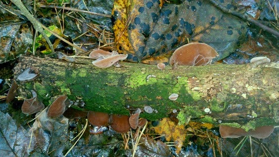
Jelly Ear Auricularia auricula-judae at Nesscliffe
Jelly Ear Auricularia auricula-judae

Also at Nesscliffe I found a branch covered with wonderful Jelly Ear (Auricularia auricula-judae) one of my favourite fungi as it looks so much like an ear. I also found a tiny jelly fungus growing on beech mast while I was searching (unsuccessfully) for the beechmast fungus Xylaria carpophila. Another highlight was the Wood Woollyfoot (Gymnopus peronatus) which as the common name suggests has a lovely ‘woolly’ base to the stipe.

Wood Woollyfoot (Gymnopus peronatus) at Nesscliffe Wood Woollyfoot (Gymnopus peronatus)
Wood Woollyfoot (Gymnopus peronatus) in habitat and close up in the lab showing woolly base to stipe

Another old favourite is “smell it before you see it” Stinkhorn (Phallus impudicus) although we only found in the ‘egg’ stage and one old specimen, both badly eaten by slugs, missing out on in its erect magnificence.
DSC_0192
Stinkhorn (Phallus impudicus) ‘egg’

Back in the lab Irene gave a demonstration of microscopy techniques for fungi and I had a go myself, finally seeing the spores, basidia and cystidia which I had failed to find on my own. These preparations involve taking a small piece of gill, putting a drop on a stain called Congo Red on top and then squashing a coverslip gently over the top. After blotting any excess liquid the slide is examined under a high power microscope. I was hoping to show some of my own slides but unfortunately I don’t have any Congo Red at home so the photomicrographs below are courtesy of Wikipedia Commons.

2009-07-12_Basidia_colored_with_Congo_Red
Basidia stained with Congo Red, the pointy bits on top are where the spores attach

By Andreas Kunze (Own work) [CC-BY-SA-3.0 (http://creativecommons.org/licenses/by-sa/3.0)], via Wikimedia Commons)

Basidia (singular basidium) are the spore bearing structures in one of the major groups of fungi, the Basidiomycota which includes most of the species identified this weekend; the shape and number of spores is used in identification. Cystidia are sticky-outy cells found on various parts of the fungal fruit body, and the position, shape and reaction with certain chemicals are important for identification.

2007-01-24_Hymenialzystiden_mit_Kristallschopf
Cystidia
By Andreas Kunze (Own work) [CC-BY-SA-3.0 (http://creativecommons.org/licenses/by-sa/3.0)], via Wikimedia Commons)

The course finished with an assignment – identifying 15 specimens and/or photographs, it was strange to think it was my last on the course and that with my uCert and MSc complete I am for the first time in 7 years not in formal education! The two fungi forays and tuition in microscopy techniques were just what I needed to get back into fungi identification, I will be dusting off my old compound microscope and giving it another go!

Friday, 25 October 2013

Earthworm bothering in Yorkshire


One of the benefits of my MSc at the Natural History Museum was the opportunity to volunteer on fieldwork for Museum projects, such as my previous post on my trip to Scotland with the Museum Dipterists. This week I was in East Riding, Yorkshire, helping with fieldwork for the Soil Biodiversity Group on a big NERC funded project on the distribution patterns of UK earthworms in pasture and the environmental factors behind their biodiversity. This work is needed because of the lack of information on how earthworms are affected by management and climate - given their importance in ecosystem functioning this needs to be addressed. In addition to the benefits of gaining more experience in soil science fieldwork this was a much appreciated change from sitting at home looking for and applying for jobs/PhDs and to have contact with fellow scientists, which had been lacking since completing my Master’s.
Since I was passing the Museum anyway, I dropped in to help Dr David Jones, who is leading the fieldwork, to prepare and transport the equipment needed and then it was off to Kings Cross Station to meet fellow volunteers Georgie and Veronika. Our destination was Hull and then onwards to the village of Patrington, were we checked into the beautiful Dunedin Hotel – it even had a dinner gong!

Dunedin Hotel (2) Dunedin breakfast room and gong

On Monday we set off for our first sampling session at the survey site – Hodgson’s Fields in Skeffling, but unfortunately the rain became very heavy and we only managed six samples. Although rather wet the group took the opportunity to visit Spurn Head, a long finger of sand (spit) stretching across the mouth of the River Humber and an important nature reserve. Myself and David braved the rain to walk a little way along the spit and see where the sea had recently breached the road.

Spurn Point road breach
Sand washed over the road that could recently be driven down to the end of the spit
Spurn Point in the rain (5)
Mudflats at Spurn Head inundated by sand

This was a trip down memory lane to my Geography A Level were quite a lot of the syllabus was on spit formation and coastal erosion. Despite being wet through and starting to feel cold I was a bit overenthusiastic and had to be retrieved by David so we could all go back to Dunedin and dry off.

Dr David, stone hunter
David collecting stones in the pouring rain, one of the last photographs I took before my camera expired. I also wrecked by waterproof over trousers by running up a dune too vigorously but gaffer tape is a wonderful thing.
Frustratingly Tuesday was more of the same but we were joined by another volunteer, Fez, which sped up the work. Sampling earthworms in grassland is a little different from the sampling I did for my MSc project. After marking out the samples using random coordinates, location and environmental measures are taken before a pit of a set size is dug from the ground. This soil is weighed and then comes the best bit – finding the earthworms! 
Skeffling by V
David and volunteers sampling earthworms. Left to right: me writing out the labels for worm tubes, Georgie with the flags used to mark the sample sites and Fez ready to weigh the soil (thanks to Veronika for the photograph)
Two methods are used to sample earthworms, which reflects the different habits (or ecoytpes) of the various species. Anecic earthworms make permanent vertical burrows deep in the soil, emerging at night to feed on leaves which they pull down into their burrows. This type of worm are forced from the soil using an irritating chemical called an expellant, we used mustard powder dissolved in water. While the pit is watched carefully for any worms that emerge a team work through the clod of soil for smaller worms which live amongst the roots, called endogeic worms. Finding these requires pulling apart the roots of the grass as they often live in-between, which was hard work when the grass was dense.

SAM_2393
Veronika (left) and myself searching for earthworms while watching the mustard pit (thanks to Georgie for photograph).
Dr David with big worm
David with a large Lumbricus earthworm from the mustard pit (thanks to Fez for the photograph)
While the earthworms are being collected and preserved in ethanol for identification back at the museum David takes a soil core which will be analysed by a team in York to measure dry weight, soil moisture and nutrient content. This involves a hamming a metal ring like a cookie cutter into the soil, often accompanied by swearing. The ring cuts a core of a set volume so each sample can be compared. Another soil sample is taken from the sorted soil which will be analysed to determine how much microbe activity there is in the sample.

Rain finally stopped play again at midday and with only one field complete out of three progress was becoming a concern but we cheered ourselves up after drying off with a lovely lunch at the excellent Sally Brown's Tea Room in Patrington.

To great relief the weather on Wednesday was much improved so we packed lunch for a full day in the field. Field two was large and tussocky, which made it hard work to travel across, especially with heavy bags full of bottles of mustard solution and soil samples. We worked under the watchful eye of sheep.

Entering field 2
Entering field number two

With field two complete it was on to the next, with the weather being much more fieldwork friendly. However this field was grazed by a herd of English Longhorn cattle, and although we had been informed they were docile, they are a large and intimidating breed. Eventually we got the hang of how to move them by shouting and walking towards them to move them out of the way so we could continue working, but it had lost us some time.

Field 3 cows (6)
I particularly like the cow second on the left which had one horn growing upwards and the other growing downwards!
Dr David, cow herder (2)

Thankfully we had excellent weather for our final day and managed to herd the cattle and confine them to a another field so it was completed. With still another field to sample another team may have to come back but for us it was off to catch the train back to London after posting the soil samples to the York laboratory.

Despite getting wet through twice in as many days I had a fantastic time and feel energised by the fieldwork and interaction with others after feeling rather isolated since my MSc finished. It was also a good opportunity to speak to others at different stages in science careers to gain tips on job applications, useful organisations, contacts and generally ‘talk science’. As a result of a conversation on the trip I am attending the British Ecological Society Mastering Ecology Symposium to present my MSc work, something I which I have not had the confidence to do before.

For more information on earthworms visit the Earthworm Society of Britain. I will be back for more earthworm bothering next month in a different county!

Monday, 7 October 2013

Chasing flies in Scotland


Between handing in my MSc thesis and my viva voce, I was fortunate enough to have the opportunity to join Dipterists from the Natural History Museum on a collection trip to Scotland as part of the Dipterists Forum Autumn Field Meeting. Despite the daunting prospect of a long journey and sharing a cottage with people I barely knew I couldn’t pass up the opportunity for an intensive week studying flies which was also my first trip across the border.
I had a fantastic time, proving to me yet again that no matter how scary something seems it is always worth doing it anyway. I even had the honour of writing up the trip for Erica McAlister’s Curator of Diptera’s Blog, so rather than blog the trip here is
Field trips through the eyes of innocents….

Dipterists under the (double) rainbow, Knappach Cottage, Kingussie
IMG_2128

Saturday, 10 August 2013

Balcony beasties – cabbage whites and their enemies


It is true you do not have to go far to see wildlife, even in the midst of a big city. Invertebrate scholars are particularly fortunate in this regard as insects, arachnids etc. really are everywhere, in your home and even on you. Here in my student room in Earls Court, London I have a little balcony which I have used to grow salad and tomatoes. Being an entomologist I soon became more interested in the creatures eating my vegetables (and eating them!) than the produce.

I watched a large white (Pieris brassicae) lay clusters of eggs on my kale a couple of weeks ago. Now if i was a ‘proper’ gardener I would remove these but as an entomologist I take pleasure in watching them develop so don’t begrudge them my salad. The caterpillars hatched and grew rapidly, first feeding together then spreading out on the container of plants. I later found a single small white (Pieris rapae) caterpillar which I had missed, as these are laid singly and must have been overlooked.

Large White caterpillars feeding on my kale on the balcony
cats
Small White caterpillar (note the leaf mines on the kale, I will discuss these in another post)
small white
By early August they were ready to pupate but I have only found on successful pupae, the rest being devoured inside out by the braconid wasp Cotesia glomerata.

The only successful large white pupa
large white pupa
All but one of the infected caterpillars mostly moved away from the food plant, climbing up the balcony to find a place to pupate, instead the braconid larvae emerged from the caterpillars and spun silk cocoons, the moribund caterpillar remaining attached and hunched over the cocoons. The caterpillars were still alive for sometime after the parasitoid larvae had emerged and pupated.

Cotesia glomerata larvae emerging from large white caterpillar, this one remained on the food plant.
IMG_1920
Caterpillar on the side of the balcony with Cotesia glomerata in their silk cocoons underneath.
IMG_1904
The small white caterpillar had a different fate, though no less gruesome. I woke up one morning to find it being macerated and carried of piecemeal by a wasp. Amongst the remains it could be seen it was also infested by Cotesia glomerata larvae.

wasp dinner
The next balcony bugs post will focus on the flies in my vegetable garden.

A weekend of hoverflies


This weekend I was at the Field Studies Centre at Preston Montford in Shropshire to learn about hoverflies. This is the penultimate course in my University Certificate in Biological Recording and Species Identification run by Manchester Metropolitan University. The course is run by Roger Morris and Stuart Ball of the Hoverfly Recording Scheme and I had previously attended their 2 day course which I enjoyed very much so I came back for more!

The course started with an introduction to hoverflies and how to recognise them, Roger and Stuart always emphasise the importance of understanding the ecology of larvae, which are often understudied in invertebrate groups. Hoverflies have particularly diverse larval biologies, with examples from all modes of life except parasitism and unusually for flies are relatively well studied, with the larvae of around two thirds of the UK's 282 species known; there is even an identification guide on them, now sadly out-of-print - the Colour Guide to Hoverfly Larvae: Diptera, Syrphidae by Graham Rotheray.

The next day began by going through some of the the keys and learning the characteristics of the different tribes, good revision for me and I am finally able to find my way around wing venation.

A hoverfly wing - R-M is your friend


Based on © Hans Hillewaert / CC-BY-SA-3.0 via Wikimedia Commons

You see the first time I came across wing venation in a key I found it very confusing, the wings just seemed to have a mass of different veins branching all over the place and trying to work out which were which was tricky. In hoverflies at least, a useful tip is to find the radial-medial (or R-M) cross vein, which is quite prominent in the middle of the wing and crosses the vena spuria or false vein (actually a deep fold), particular to hoverflies. This immediately locates the radial 4 + 5 vein which runs along the top, the discal cell which lies underneath and the first basal cell which it forms the outer border of. Now venation is not so daunting!

On the Sunday we were allowed out of the lab to see what hoverflies could be found around Preston Montford field centre which were then brought back for killing, pinning and identification. A few other diptera groups sneaked in as well!

My modest catch, well I had a MSc thesis to write!


The final day comprised a test towards the uCert qualification which I passed. Hopefully I will also pass the 2nd assignment and then I am just one more identification course away from the Certificate!

Thanks to Roger and Stuart for another expertly run course!